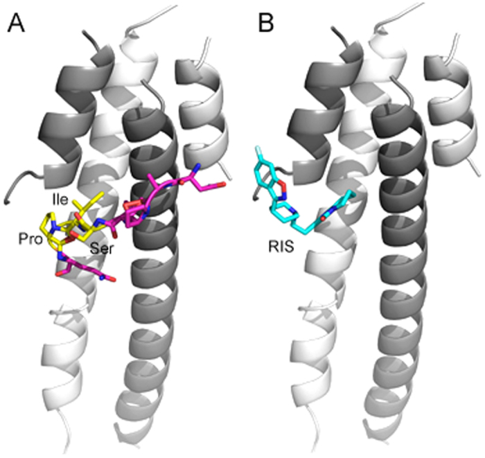
Figure 1. NAP and RIS binding prediction to EB3.

(A) NAP (NAPVSIPQ) peptide binding to EB3. EB3 dimer is shown as white and gray cartoons (PDB 3TQ7, residues in chain A 197–248 and chain B 204–258). The peptide is shown as purple sticks, while the SIP motif is colored in yellow. The binding orientation was predicted using structural alignment of EB3 (PDB 3TQ7) to EB1-EB3 complex with peptide MACF (PDB 3GJO). (B) RIS binding to EB3, as predicted by Swissdock. RIS is shown in cyan sticks. The figure was generated using Pymol.
