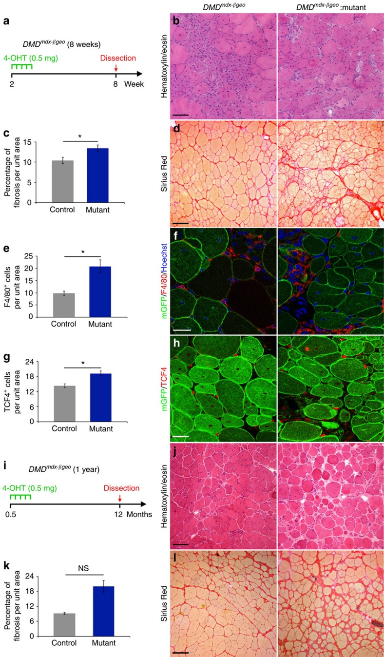
Figure 2. Loss of Numb/Numbl exacerbates the dystrophic muscle phenotype.
Data were collected from transverse TA muscle cryosections from DMDmdx-βgeo mice. (a) Scheme of the experiments. (b) Histological staining with H&E. (c) Quantification of fibrosis in mutants and controls in 8-week-old DMDmdx-βgeo mice based on Sirius Red histology (control: n=8 TA, 5 mice, 10.4±0.8% fibrosis per unit area; mutant: n=13 TA, 7 mice, 13.5±0.8% fibrosis per unit area; MW P=0.0303). (d) Histological staining with Sirius Red. (e) Quantification of F4/80+ cells (control: n=4 mice, 9.8±0.9 cells per unit area; mutant: n=5 mice, 20.8±2.7 cells per unit area; MW P=0.0159). (f) Immunofluorescence staining for macrophages using anti-F4/80 antibody. (g) Quantification of Tcf4+ (control: n=4 mice, 14.3±0.7 cells per unit area; mutant: n=4 mice, 19.2±1.0 cells per unit area; MW P=0.0286). (h) Immunofluorescence staining for fibroblasts using anti-Tcf4 antibody. (i) Scheme of the experiments. (j) Histological staining with H&E. (k) Quantification of fibrosis in mutants and controls in 1-year-old DMDmdx-βgeo mice based on Sirius Red histology and (control: n=3 TA, 3 mice, 9.1±0.4% fibrosis per unit area; mutant: n=3 TA, 3 mice, 20.1±2.4% fibrosis per unit area; MW P=0.10). (l) Histological staining with Sirius Red. Quantifications are presented as mean±s.e.m.. Scale bars b,d,j,l: 100 μM; f,h: 25 μM. NS, not significant.

