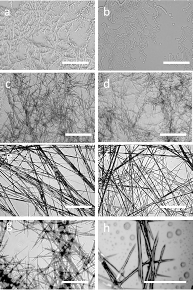
Figure 3. Optical microscopy images of the assemblies formed by sonication.
Optical microscopy images of xerogels formed by (a) Peptide 1 in hexane-ethyl acetate (3:1 v/v) and (b) Peptide 1 in toluene; (c) Peptide 2 in hexane-ethyl acetate (3:1 v/v), and (d) Peptide 2 in toluene; (e) Peptide 3 in hexane-ethyl acetate (3:1 v/v), and (f) Peptide 3 in toluene; (g,h) representative images of sonication-induced self-assembled aggregates formed by peptide 4 in hexane-ethyl acetate (3:1 v/v) and toluene, respectively. Scale bars represent 20 μm.

