Figure 2.
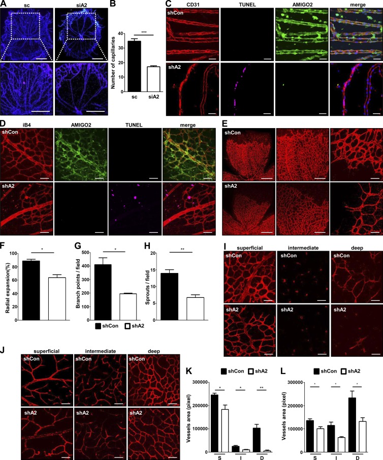
AMIGO2 is required for angiogenesis in vivo. P3.5 mice were injected with Amigo2-specific or control siRNA and shRNA, and experiments were performed at P5.5, P8.5, and P15.5. (A) Impairment of hyaloid vessel structures in Amigo2 siRNA–injected mice. Bars, 200 µm. (B) Capillary quantification. n = 5. (C) Hyaloid vessel staining with CD31, AMIGO2, and TUNEL was performed in Amigo2 shRNA–injected P5.5 mice. (D and E) Visualization of blood vessels by iB4 staining with AMIGO2 and TUNEL staining of Amigo2-depleted retinas from P5.5 mice. Bars: (D) 50 µm; (E, left to right) 500 µm, 200 µm, and 50 µm. (E) Middle and right images are enlargements of the left image. (F–H) Radial expansions, retinal vascular branch points, and sprouts per field were quantified. n = 5. (I–L) Immunostaining of blood vessels by iB4-staining of Amigo2 shRNA–injected P8.5 retinas (I and K) and P15.5 retinas (J and L). Superficial (S), intermediate (I), and deep (D) layers of retinal vessels were taken and quantified. Bars, 50 µm. n = 5. Data were collected from mice (n = 5–6) and analyzed using a two-tailed unpaired t test. *, P < 0.05; **, P < 0.005; ***, P < 0.0001. Data are means ± SD. shCon, control shRNA; shA2, Amigo2 shRNA.

