Figure 5.
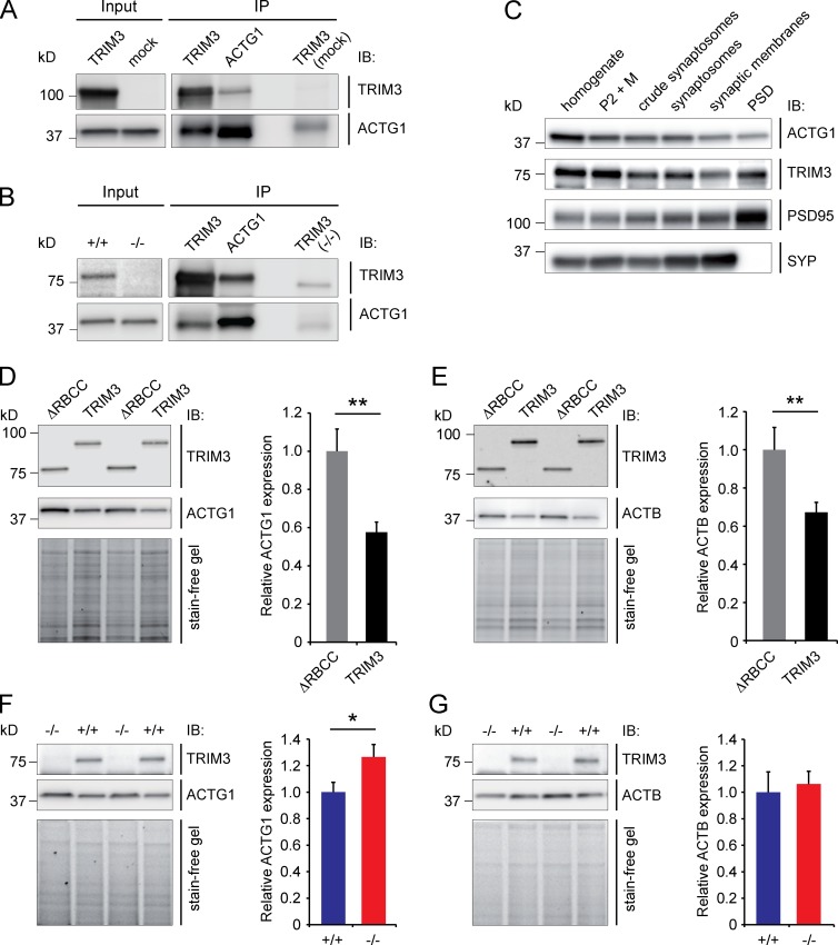
TRIM3 interacts with ACTG1 and regulates ACTG1 levels. (A) HEK293 cells were transfected with TRIM3 or mock transfected. TRIM3 and ACTG1 were immunoprecipitated (IP) from the lysates, immunoblotted and stained for TRIM3 and ACTG1. TRIM3 can be observed in the ACTG1 IP and ACTG1 in the TRIM3 IP. No or significantly less TRIM3 or ACTG1 protein was detected in IPs on the mock-transfected cell lysate. (B) TRIM3 and ACTG1 were immunoprecipitated from hippocampal synapse-enriched fractions, immunoblotted, and stained for TRIM3 and ACTG1. TRIM3 can be observed the ACTG1 IP and ACTG1 in the TRIM3 IP. No or significantly less TRIM3 or ACTG1 protein was detected in the TRIM3 IP on the Trim3−/− tissue sample. (C) ACTG1 and TRIM3 are colocalized in hippocampal synapses. Biochemical fractionation of hippocampal tissue followed by immunoblot analysis showed that ACTG1 and TRIM3 are both detected in synaptic fractions. Enrichment of synapses is indicated by PSD-95 and SYP staining. (D and E) TRIM3 regulates actin levels in HEK293 cells. HEK293 cells were transfected with TRIM3 or ΔRBCC-TRIM3. Lysates were immunoblotted and stained for TRIM3 and ACTG1 (D) or TRIM3 and ACTB (E). Normalized protein levels of both ACTG1 and ACTB were significantly reduced in TRIM3-expressing cells compared with cells expressing ΔRBCC-TRIM3 (means ± SEM, two-tailed t test, **, P < 0.01, n = 8 cultures per condition. (F and G) TRIM3 regulates ACTG1 levels, but not ACTB levels, in hippocampal neurons. Cultured hippocampal neurons were lysed, immunoblotted, and stained for TRIM3 and ACTG1 (F) or TRIM3 and ACTB (G). Normalized protein levels of ACTG1, but not ACTB, were significantly higher in neurons from Trim3−/− mice compared with wild-type control neurons (means ± SEM, two-tailed t test, *, P < 0.05, n = 8 cultures per genotype).

