Figure 7.
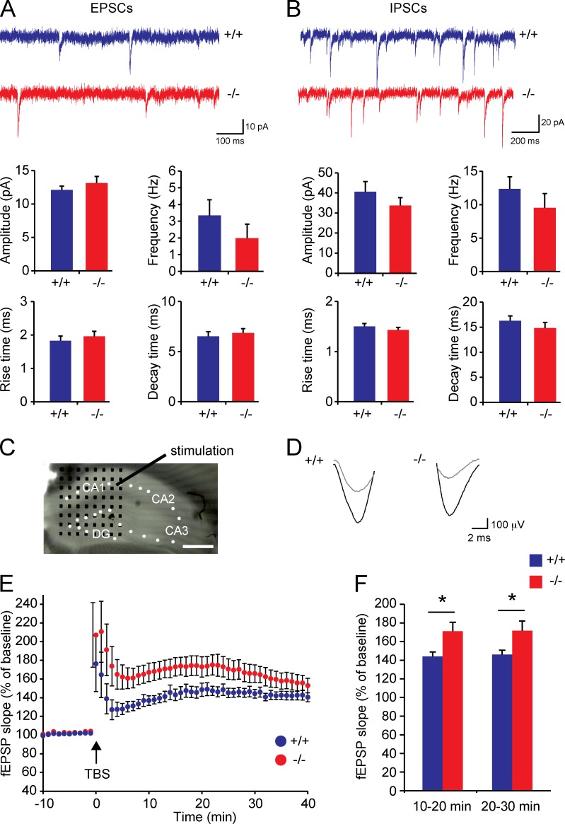
Hippocampal LTP is increased in Trim3−/− mice. (A and B) Spontaneous excitatory (A) and inhibitory (B) synaptic transmission are unaffected in Trim3−/− mice. Excitatory synaptic currents (mEPSC) and inhibitory synaptic currents (mIPSCs) were recorded from hippocampal slices. Frequency, amplitude, rise time, and decay time of both excitatory and inhibitory currents were unaffected in Trim3−/− mice compared with wild-type controls (means ± SEM, n = 6–8 for EPSCs, n = 10 for IPSCs). (C) LTP was induced in acute hippocampal slices using a single stimulation electrode in the Schaffer collateral pathway, and field excitatory postsynaptic potentials (fEPSPs) were recorded using an 8 × 8 multielectrode array (black dots) in the CA1 area. Bar, 400 µm. (D) Example field EPSP traces from a Trim3−/− mice and a wild-type control before (gray) and after (black) tetanus stimulation indicate stronger potentiation in Trim3−/− mice. (E) Averaged fEPSP data show enhanced LTP in Trim3−/− animals, in particular in the first 30 min after theta burst stimulation (TBS). (F) Quantification of the mean amount of potentiation at 10–20 min and 20–30 min after TBS show a significant increase in Trim3−/− mice (means ± SEM, two-tailed t test, *, P < 0.05, n = 15/16 slices obtained from 9/10 animals per genotype).

