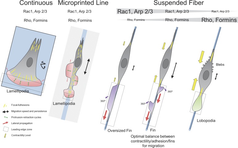
Figure 10.
Schematic representation. On continuous substrate, the leading edge (dashed black box) is narrow in the axis of migration and determined by lamellipodia width and is very large in the transverse axis. Protrusion-retraction cycles of this region, along the axis of migration, promote cell motion, whereas lateral diffusion of the lamellipodia (red arrows) does not assist in migration. Migration is slow and poorly persistent (black track). On microprinted lines, the leading edge of the cell is now restricted by the width of the line, and short protrusion-retraction cycles in this zone are responsible for leading edge advancement. Focal adhesions are distributed over the entire cell length, resulting in the nucleation of new lamellipodia sites that do not promote effective migration. The lateral propagation could help migration, but the supportive nature of the substrate around the lines promotes lateral extension rather than propagation. Migration is fast and moderately persistent as the cell can easily nucleate new leading edge at the back of the cell. On suspended fibers, the leading edge is long in the axis of migration but narrow in the other axis. The lamellipodia-like actin under Rac1-Arp2/3 signaling cascade forms fin-like protrusions at the focal adhesion site. The lateral diffusion of fins is the leading cause for edge extension and cell migration. No protrusion retraction cycles are observed, as the fins are moving forward or backward. The fins are rotating and capable of changing direction and polarity once reaching the extreme end of the leading edge. Migration is fast and highly persistent. There is an optimal balance between contractility, adhesion and fins to promote the migration (contribution of contractility showed by yellow arrows). The Rho-formin pathway is important for fin creation but does not participate in their propagation. If the Rac1-Arp2/3 pathway becomes predominant, the cell shifts behavior and harbors oversized fins. If the Rho-formin pathway becomes predominant, the cell shifts behavior and harbors large lobopodia protruding at the front and lateral blebs at the back. Those cells are poorly persistent as lobopodia can change direction, in part because of strong hydrostatic pressure in the cell.

