Figure 2.
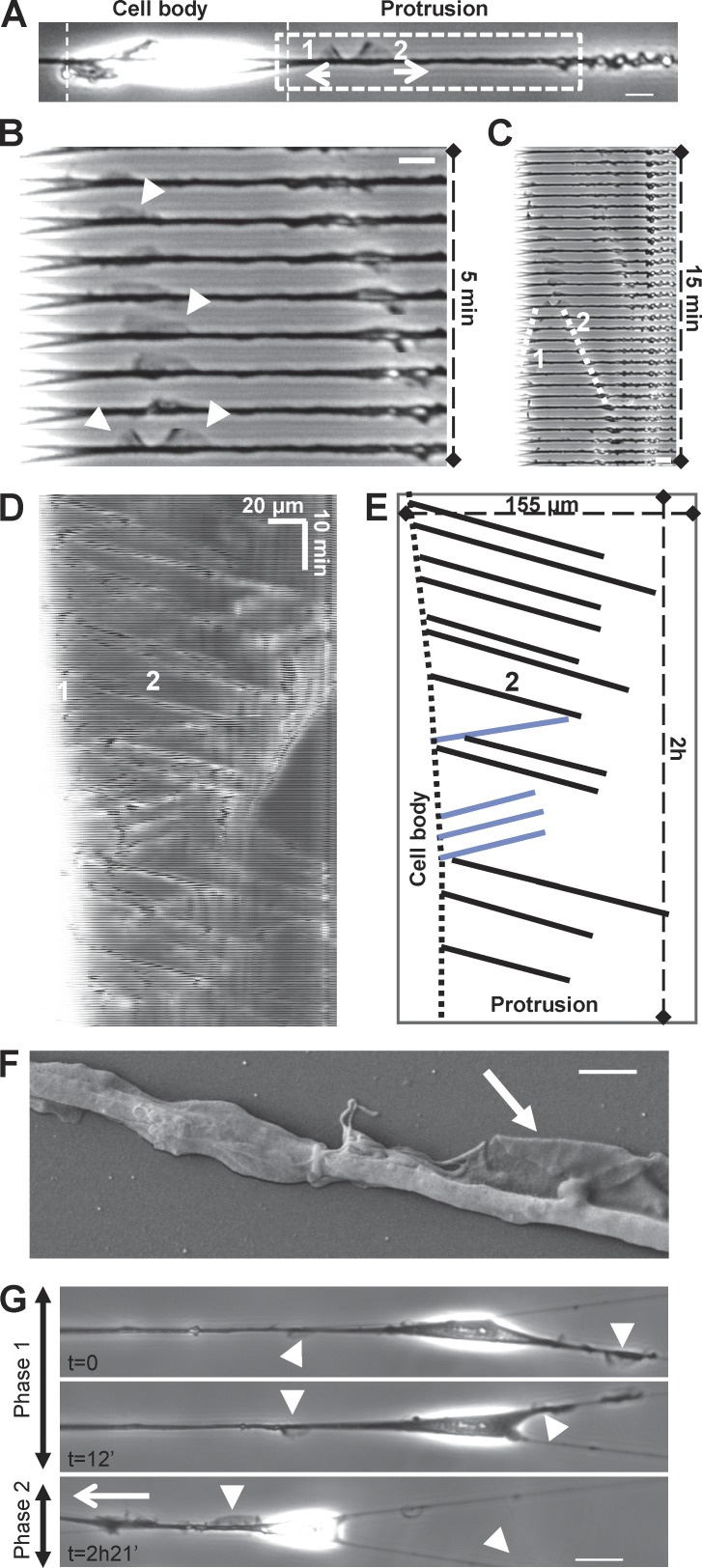
Fibroblasts display cyclical “fin-like protrusion” on nanofibers. (A) 3T3 fibroblast on fiber presenting two fin-like protrusions. Bar, 10 µm. (B and C) Time-sequenced zooms of the leading edge during fin propagation. Bar, 10 µm. (D) Kymograph of fin motion along the fiber. The white zone (left) corresponds to the end of the cell body where fins are born before propagating. (E) Schematic analysis of kymograph (D) showing properties of fin-like protrusion including robust speed and propagation between cell body and cell tip. Black lines, forward-moving fins; blue lines, backward-moving fins. (F) Scanning electronic microscopic image of the fin-like protrusion (arrow). Bar, 2 µm. (G) 3T3 in a multiple fiber situation. Two phases: phase 1, no net migration with fins on both fibers; phase 2, fins only in one fiber leading to the migration of the cell along this fiber. Arrowheads, fin-like protrusions; white arrow, direction of migration. Bar, 20 µm.

