Figure 5.
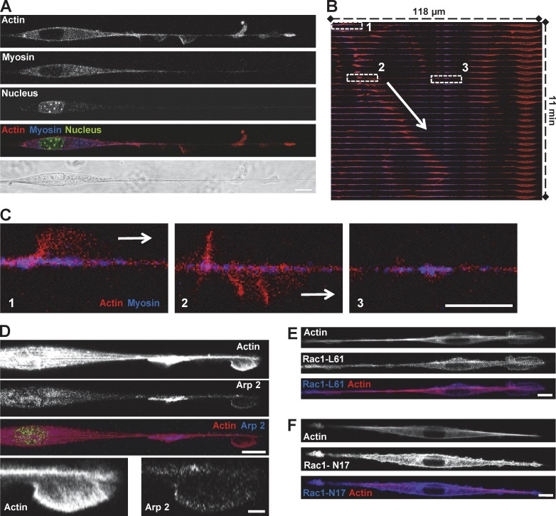
Fin-like protrusion is driven by Arp2/3 mediated actin polymerization. (A–C) 3T3 cells transfected with GFP-Actin and RFP-MLC on fiber. Bar, 10 µm. (A) Fins contained actin whereas myosin was concentrated in the cell body. Bar, 10 µm. (B) Kymograph of the merge color (actin in red, myosin in blue) showing propagation of the fin (arrow). (C) 3-Zoom image from B. 1, fin; 2, same fin from 1 after turning around the fiber; 3, myosin patches on fiber. Bars, 10 µm. (D) 3T3 cells were transfected with GFP-Actin and mCherry-Arp2/3 on fiber. Bars: (main) 10 µm; (zooms) 2 µm. (E and F) 3T3 cells were transfected with YFP-Rac1-L61 (Rac dominant positive, oversized fins) or YFP-Rac1-N17 (Rac dominant negative, no fin-like protrusion) and RFP-Actin and plated on fiber. Bars, 10 µm.

