Figure 2.
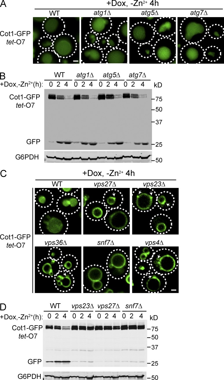
ESCRTs, but not the autophagy machinery, are required for the degradation of Cot1-GFP. (A) Localization of tet-O7 Cot1-GFP in WT and autophagy mutants 4 h after Zn2+ withdrawal from YNB media and addition of 2 µg/ml Dox. (B) Degradation kinetics for tet-O7 Cot1-GFP in WT and autophagy mutants. G6PDH was used as a loading control. Same volume of cells was loaded, with 1 OD600 cells loaded at 0 h. (C) Localization of tet-O7 Cot1-GFP 4 h after Zn2+ withdrawal in WT and ESCRT mutants. Arrows highlight the aberrant endosomes. (D) Degradation kinetics for tet-O7 Cot1-GFP in WT and ESCRT mutants. G6PDH was used as a loading control. Same volume of cells was loaded, with 1 OD600 cells loaded at 0 h. Bar, 1 µm.

