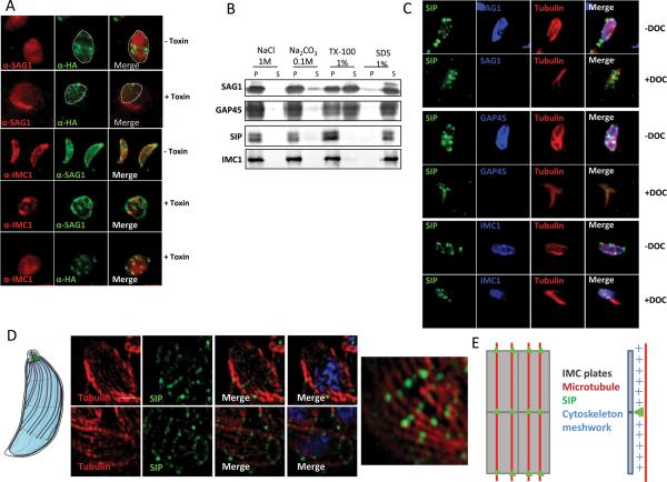
Figure 3. TgSIP is associated with the detergent resistant skeleton of the parasite pellicle.
A. Incubation of TgSIP-HA3 parasites with aerolysin alpha-toxin revealed that TgSIP is associated with the IMC. In untreated cells, the transversal lines of TgSIP occupied the width of the parasite labeled with the plasma membrane marker SAG1. Following alpha-toxin treatment, the dramatic swelling of the plasma membrane separates it from the underlying IMC. In this condition, TgSIP followed the IMC1 staining and became distant from the plasma membrane, demonstrating the association of TgSIP with the IMC.
B. Detergent extraction profile of TgSIP after solubilisation of HA-tagged TgSIP parasites with 1% Triton X-100, 1% SDS, 1M NaCl or 0.1M Na2CO3. Extracts were subjected to SDS-PAGE, blotted and probed with antibodies as indicated. As expected, the GPI-anchored plasma membrane SAG1 and the acylated-GAP45 protein were solubilized in 1% Triton X-100. In contrast, the detergent resistant IMC protein meshwork containing IMC1 remained in the pellet upon Triton-X-100 solubilization. As IMC1, TgSIP was only solubilized in 1% SDS, demonstrating that it is probably embedded in the meshwork of the IMC.
C. Localization by IFA of TgSIP after DOC-extraction. As expected, in DOC-extracted parasites, SAG1 and GAP45 were not detected by IFA, while the tubulin and IMC1 proteins remained in the cytoskeleton ghost. IFA with anti-HA revealed the presence of dots of SIPHA3 associated with the cytoskeleton ghost, indicating that TgSIP associates with the IMC network.
D. Structured illumination Super Resolution (SIM-SR) microscopy of parasites using anti-HA and anti-tubulin antibodies. Scale bars represent 1 μm. Upper panel: single plane of a Z-stack acquisition. Lower panel and right panel: maximum intensity projection. The nuclei were stained with DAPI (blue). TgSIP showed a periodic row organization matching in most cases the periodicity of one of the microtubules. On the left, schematic representation of the IMC structure and of the subpellicular microtubules of Toxoplasma tachyzoites.
E. Schematic representation of TgSIP localization on IMC sutures. The plates are represented by rectangles and the subpellicular microtubules are in red; TgSIP is indicated by green dots located on the transverse sutures at the intersections with microtubules; lateral view (right) shows that TgSIP is located in the subpellicular network.

