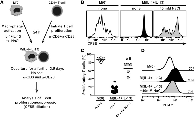
Figure 4. High salt reduces the suppressive capacity of M(IL-4+IL-13) macrophages.
(A) Schematic of the in vitro coculture assay. Briefly, macrophages were activated into M(IL-4+IL-13) with/without an additional 40 mM NaCl for 24 hours. Excess salt was then removed by repeated washing of the cells. Coculture of CFSE-labeled CD4+ T cells (from whole splenocytes) and preactivated macrophages occurred in the presence of plate-bound α-CD3 and α-CD28, but without any additional salt. CD4+ T cell proliferation was monitored by flow cytometry. (B) CFSE fluorescence histograms of gated CD4+ T cells incubated with macrophages at a ratio of 6:1 (T cell/macrophage). Macrophages were unactivated (M[0]) or activated with IL-4+IL-13 in the absence (none) or with high salt. (C) Quantification of B. Bars show the mean percentage of proliferating CD4+ T cells (n = 5 technical replicates). The experiment was repeated at least 3 times independently. *P < 0.05 vs. M(0) and #P < 0.05 vs. M(IL-4+IL-13) none by 1-way ANOVA. (D) The level of PD-L2 expression of macrophages from B was determined by flow cytometry. Numbers above each line indicate the mean fluorescence intensity. A similar trend was observed in 3 independent experiments.

