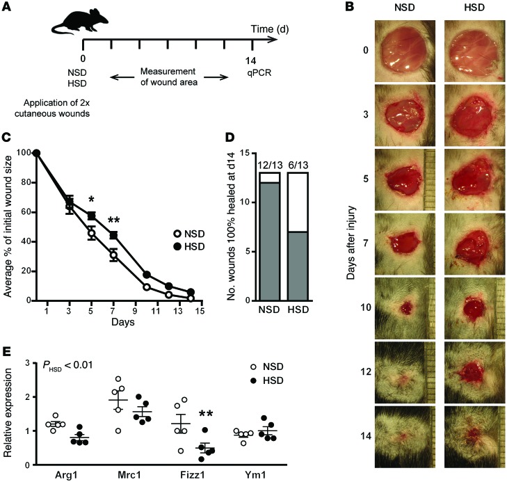
Figure 6. HSD leads to impaired wound healing in vivo.
(A) Two cutaneous wounds were applied to the back of mice, which were then fed an NSD or HSD for 14 days. The closure of the wounds was monitored at the desired times in this 14-day period, at the end of which skin samples from the wounded area were subjected to qPCR analysis. (B) Representative images of wounds from mice on an NSD and HSD during 14 days after wounding. (C) The percent change in wound area is plotted over time. The experiment was repeated twice independently, at which point the values from 7 individual mice were pooled (n = 13 wounds per group). *P < 0.05 and **P < 0.01 by 2-way ANOVA. (D) The number of wounds completely healed (gray) vs. incompletely healed (white) at the end of the experiment (14 days). (E) Real-time qPCR analysis of M2 signature genes at wound sites from mice on an NSD and HSD. The P value shown for the effect of HSD was calculated by 2-way ANOVA. **P < 0.01. n = 5 (biological).

