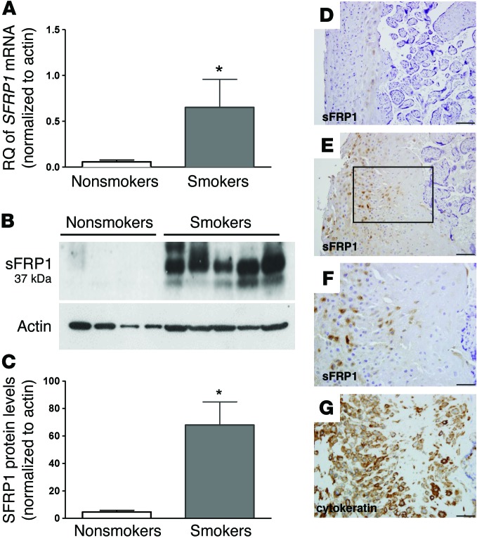
Figure 1. Elevated SFRP1 gene and protein expression in the placentas of smokers.
(A) Relative quantification (RQ) of SFRP1 mRNA levels measured, as by qRT-PCR of placentas from smoking mothers (n = 7) and nonsmoking mothers (n = 14) (mean ± SEM, *P < 0.05 versus nonsmoking controls by t test). (B) Representative immunoblot of sFRP1 and β-actin protein expression in placental extracts obtained from nonsmoking and smoking mothers. (C) Quantification of immunoblots by densitometry of sFRP1 and β-actin protein expression in placental extracts obtained from nonsmoking (n = 8) and smoking (n = 9) mothers (mean ± SEM, *P < 0.001 versus nonsmoking controls by t test). (D–G) Representative images of human placental tissue sections with immunostaining for anti-sFRP1 from (D) a nonsmoker and (E) a smoker. Serial placental sections from a smoker with immunostaining (F) for anti-sFRP1 and (G) for anti-cytokeratin show localization of sFRP1 to the extravillous trophoblast. The boxed region in E is shown at high magnification in F and G. Five placentas from smokers and five placentas from nonsmokers were compared. Scale bar: 100 μm (D and E); 50 μm (F and G).

