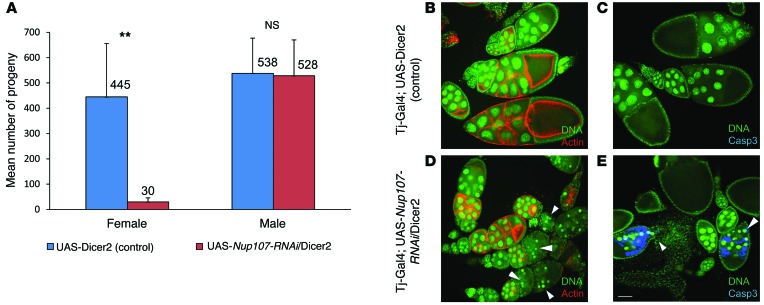
Figure 2. Gonadal somatic cell knockdown of Nup107 in Drosophila.
(A) Reduced total number of progeny in female Nup107-RNAi knockdown flies. Mean number of progeny in UAS-Nup107-RNAi/UAS-dicer2 flies compared with that in control UAS-Dicer2 female or male flies, expressed with the gonadal somatic–specific Tj-Gal4 driver. Nup107 knockdown in female flies results in 15-fold fewer progeny. Nup107 knockdown in male flies does not affect the number of progeny. The mean number of progeny in >10 flies in each of 4 independent experiments is indicated above the bars. **P = 0.007, 2-tailed, unpaired Student’s t test. (B–E) Structural defects and apoptosis in ovarioles of Nup107-RNAi knockdown and control flies. (B and C) Tj-Gal4; UAS-dicer2 control ovarioles. Control egg chambers. DNA in the nurse cells appears normal (green), (B) overall egg chamber structure is intact (normal actin staining [red]), and (C) no apoptosis is observed (no caspase-3 staining [blue]). (D and E) Tj-Gal4; UAS-Nup107-RNAi/UAS-dicer2 ovarioles. Egg chambers in Nup107-RNAi knockdown flies, expressed using the somatic gonadal-specific tj-Gal4 driver. (D and E) DNA in the nurse cells appears condensed and punctate (arrowheads). (D) Disintegration of the egg chamber structure is observed around these condensed nuclei (no actin staining), and (E) apoptosis is apparent (caspase-3 staining). DNA was stained by Sytox (green), actin was stained by rhodamine phalloidin (red), and apoptosis was identified using anti-cleaved caspase-3 (casp-3) staining (blue). Scale bar: 100 μm.

