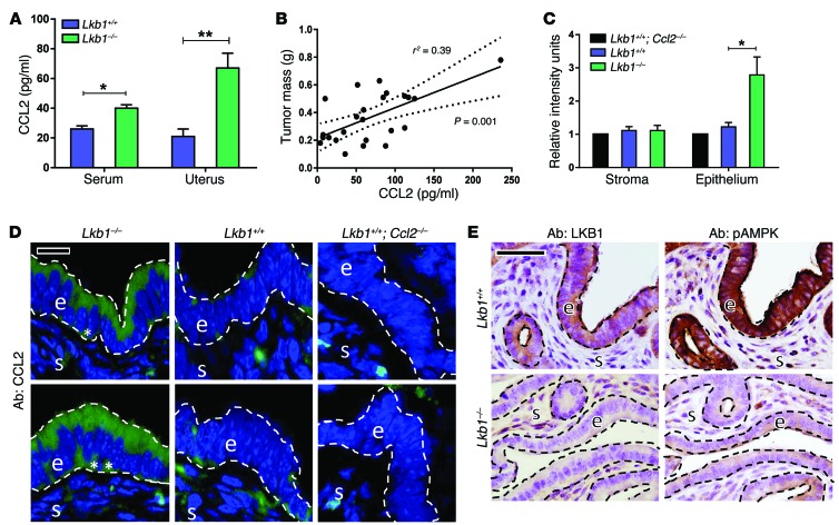
Figure 3. Conditional Lkb1 knockout in murine endometrial epithelium results in endometrial cancers characterized by high CCL2 production.
All experiments were conducted in conditional knockout Lkb1–/– and sibling control Lkb1+/+ female mice at 12 weeks of age, the time point at which myometrial invasion first occurs in this well-characterized model. Tissues were harvested at proestrus. (A) ELISA of serum or uterine protein lysates showing a significant increase of CCL2 levels following Lkb1 deletion (n = 14 Lkb1+/+, n = 24 Lkb1–/–). (B) CCL2 levels in tumor lysates (per ELISA) plotted against tumor mass from Lkb1–/– animals, showing a direct correlation between tumor mass and CCL2 levels (n = 24 mice, Pearson coefficient r2 = 0.39 with P = 0.001 per 2-tailed t test). (C) CCL2 expression in endometrial epithelium by image analysis (regions analyzed correspond to those enclosed by dashed lines in the next panel). Tissue sections were stained with a validated CCL2 antibody, and green CCL2 signal quantitation was performed using ImageJ (http://imagej.nih.gov/ij/) (n = 6 animals per experiment). Expression was normalized to the background signal present in Ccl2–/–; Lkb1+/+ uterine epithelium. (D) CCL2 immunofluorescence of uterine tissue sections. s, stroma; e, epithelium. Asterisks denote basal CCL2 expression. Two different regions are shown for each genotype. (E) LKB1 and pAMPK (Thr172) immunohistochemistry of uterine tissue sections from Lkb1+/+ and Lkb1–/– mice. As expected, Lkb1 deletion in endometrial epithelium resulted in undetectable LKB1 protein as well as reduced pAMPK compared with control siblings. Statistical significance in A and C was determined by Student’s t test. *P < 0.005; **P < 0.001. Scale bars: 50 μm. Error bars = SEM.

