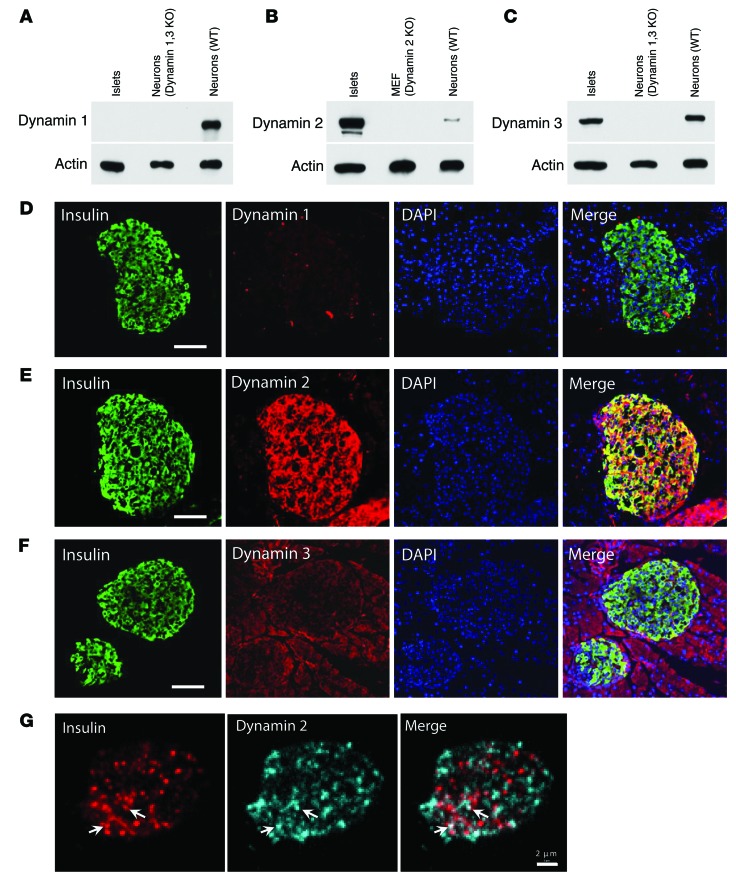
Figure 1. Dynamin isoforms in mouse pancreatic islets.
(A–C) Western blots for dynamin 1, 2, and 3 in purified mouse islet lysate (lane 1). The specificity of antibodies was verified using cell lysates with the indicated dynamin genotype as positive and negative controls (lanes 2 and 3). 110–150 islets were used for each lane. Each panel shows representative results from 3 to 5 independent experiments. MEF, mouse embryo fibroblasts. (D–F) Immunofluorescence of dynamin 1, 2, and 3 in mouse pancreatic islets. (G) Subcellular distribution of dynamin 2 in β cells (bottom optical section using spinning disk confocal microscopy views). Arrows indicate dynamin 2 puncta colocalized with insulin granules. Scale bars: 50 μm (D–F); 2 μm (G).

