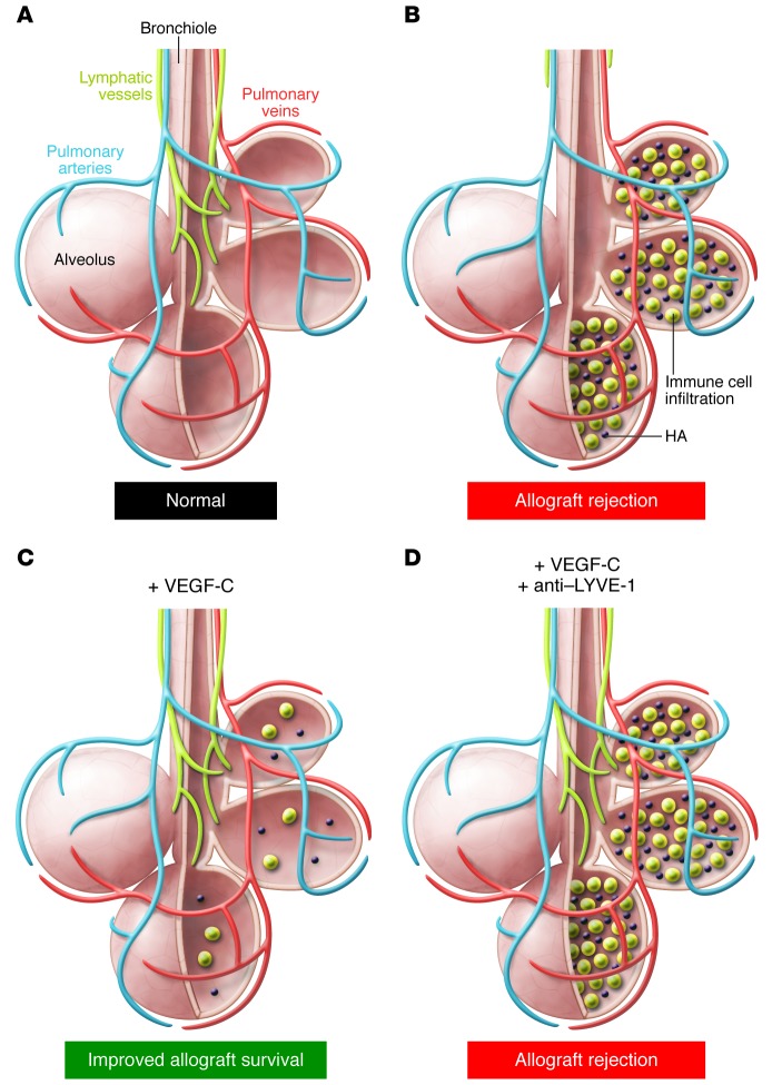
Figure 1. Lymphatic vessels improve lung allograft survival and function.
(A) Normal lung parenchyma showing air-filled alveoli, blood vessels, and lymphatic vessels that reach the alveoli along the airway. (B) During lung transplantation, the major airways and blood vessels are preserved, but lymphatic vessel density is reduced. In this issue, Cui et al. (3) demonstrate that in rejecting murine lung allografts, inflammatory cells infiltrate the alveoli and HA levels increase. (C) Animals given systemic VEGF-C, which promotes lymphangiogenesis, exhibit increased lymphatic vessel density, reduced HA accumulation, and improved allograft survival. (D) In VEGF-C–treated animals, inhibition of the interaction between HA and LYVE-1 reverses the beneficial effects of VEGF-C treatment, despite the increase in lymphatic vessel density.

