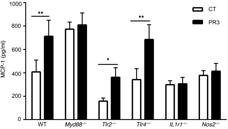
Figure 4. The proinflammatory response induced by PR3 on apoptotic cells was dependent on the IL-1R1/MyD88 signaling pathway and mediated by NO.
Thioglycolate-elicited macrophages from C57BL/6 (n = 11), Myd88–/– (n = 12), Tlr2–/– (n = 8), Tlr4–/– (n = 8), Il1r1–/– (n = 6), and Nos2–/– (n = 6) mice were cultured for 24 hours with apoptotic control or PR3-expressing cells and MCP-1 secretion was assessed in duplicate by ELISA. Values are shown as mean ± SEM. *P < 0.05; **P < 0.01. Significant differences between groups were determined by the Mann-Whitney U test.

