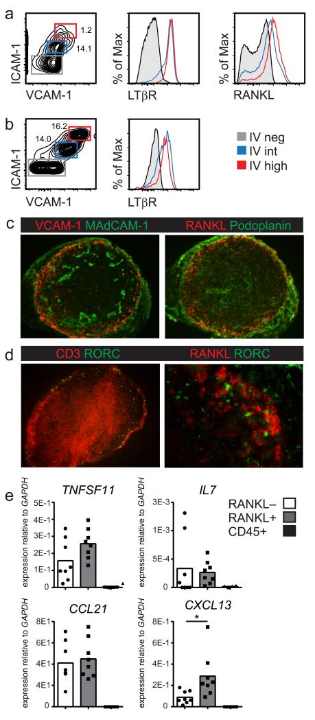
Figure 1. Stromal LTo cells in human fetal lymph nodes.
Fetal human lymph nodes and mesenteries were analyzed for presence of cells resembling murine LTo cells. (a) Expression of VCAM-1, ICAM-1, LTβR and RANKL on CD45-CD31− stromal cells from first trimester mesenteries. (n=3; age range: 8-10 weeks gestation) (b) Expression of VCAM-1, ICAM-1 and LTβR on CD45−CD31− stromal cells dissected from second trimester lymph nodes. (n=5; age range: 8-10 weeks gestation) (c) Localization of VCAM-1+ and RANKL+ stromal cells directly underneath the Podoplanin expressing subcapsular sinus (magnification 100x) (d) Co-localization of Rorγt+ cells and RANKL+ cells in fetal lymph nodes (magnification 100x/250x) (n=5) (e) Transcript analysis by qPCR of CD45−CD31− RANKL+ and RANKL− stromal cells purified from second trimester lymph nodes (n=8) compared to total CD45+ cells (n=4). (c-e: second trimester; age range 15-22 weeks gestation)

