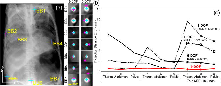
Figure 5.

(a) Example registration for the thoracic radiograph acquired with an SDD of ~1200 mm with the true and estimated position of seven target BBs overlaid. (b) Zoomed-in views of the each target BB, showing the true (cyan) and estimated (magenta) locations for the 9- and 6-DOF registration methods. (c) Median PDE (over 100 trials) for each image in the nine cases in the cadaver study (three anatomical regions and three values of SDD).
