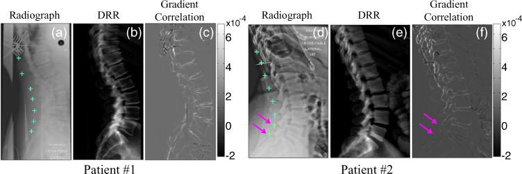
Figure 7.

9-DOF 3D–2D registration of anatomical landmarks (spinous processes) in patients undergoing spinal intervention. (a,d) Intraoperative radiographs superimposed by true (defined by a radiologist, cyan) and estimated (via 3D–2D registration, yellow) landmark locations. (b,e) DRRs computed at the registration result, shown for side-by-side comparison to the true radiograph. (c,f) GC similarity metric image at the registration solution. Overall, the registration result agreed with the true locations, with slight error (<2 mm) noted in the lumbar spine of patient #2 due to an anatomical deformation marked by pink arrows. The color-bars indicate the grayscale window of the GC image (c,f). The windows of a, b, d, e were adjusted manually.
