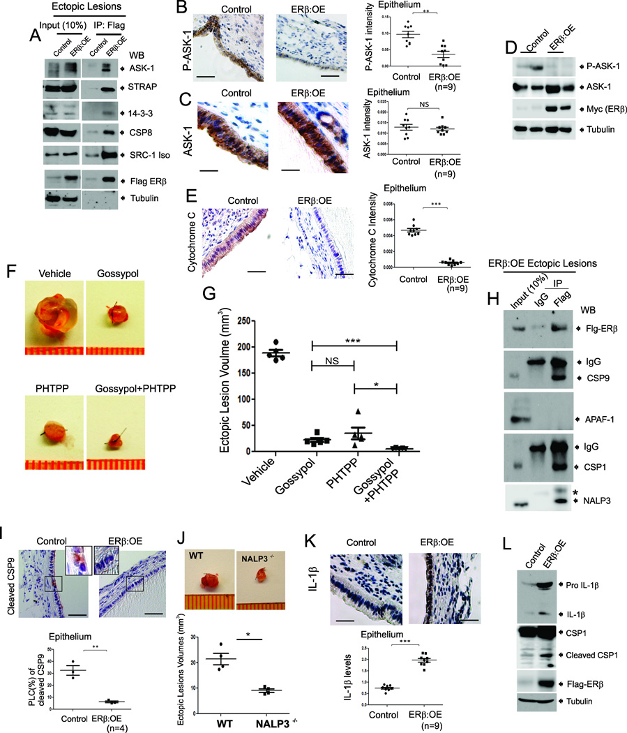
Figure 6. ERβ Interacts with TNFα-induced Apoptosis Complexes and the Inflammasome in Endometriotic Tissues of Mice with Endometriosis.
(A) Flag-ERβ complexes immunoprecipitated (IPed) with a Flag antibody from ectopic lesions of Control and ERβ:OE mice with endometriosis followed by western blotting (WB) with antibodies against ASK-1, STRAP, 14-3-3, CSP8, SRC-1, Flag and tubulin.
(B–C) IHC and quantitative analyses of phospho-Thr845-ASK-1 (P-ASK-1) (B) and total ASK-1 (C) in Control and ERβ:OE ectopic lesions.
(D) Western blot analyses of phospho- Thr845-ASK-1 (P-ASK-1), total ASK-1, ERβ and tubulin in Control and ERβ:OE ectopic lesions.
(E) IHC and quantitative analyses of cytochrome C levels in Control and ERβ:OE ectopic lesions.
(F and G) Regression of ectopic lesion growth in endometriosis-induced C57BL/6J mice subcutaneously treated with Gossypol, PHTPP or their combination compared to vehicle (F). Quantification of ectopic lesion volume in panel F is shown in the graph (G).
(H) The IPed Flag-ERβ complex from ERβ:OE ectopic lesions with a Flag antibody or IgG followed by western blotting with antibodies against Flag, CSP 9, APAF1, CSP1 and NRLP3. *, Non-Specific Protein.
(I) IHC and quantitative analyses of cleaved CSP9 levels in Control and ERβ:OE ectopic lesions. Higher magnification views of the boxed regions.
(J) Ectopic lesions isolated from C57BL/6J (WT) and NALP3−/− mice with endometriosis.
(K) IHC and quantitative analyses of IL-1β levels in Control and ERβ:OE ectopic lesions.
(L) Western blot analyses of levels of IL-1β, CSP1, Flag-tagged ERβ and tubulin (as a protein loading control) in ectopic lesions of control and ERβ:OE mice with surgically induced endometriosis. See also Figure S4 and S5.

