Fig. 1.

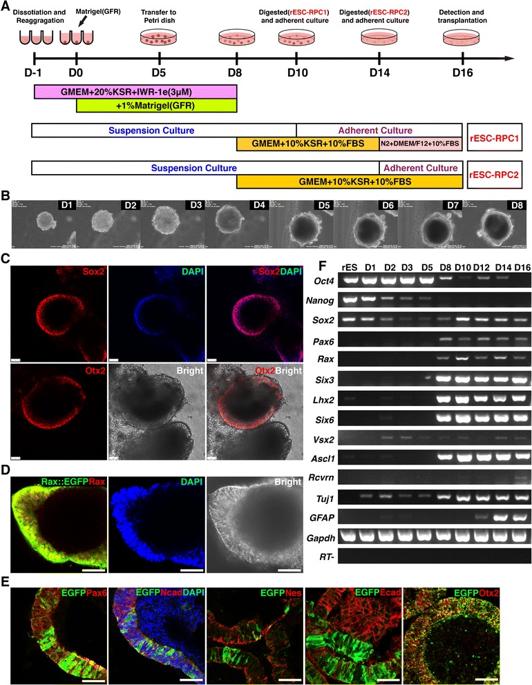
In vitro differentiation of rat ESCs into RPCs. a Schematic diagram illustrating the strategy for differentiating rESCs into RPCs in this study. b Images of morphological changes of differentiating rESCs from day 1 to the formation of neuroectoderm-like structure at day 8. c Whole mount immunofluorescence staining, using antibodies against Sox2 and Otx2 (red), and bright images of rESC-derived neuroectoderm-like structure at day 8. DAPI (blue) was used to highlight the nuclei. Scale bar: 50 μm. d Whole mount immunofluorescence staining using antibody against Rax (red) and bright images of Rax::EGFP rESC-derived neuroectoderm-like structure at day 8. DAPI (blue) was used to highlight the nuclei. Scale bar: 50 μm. e Immunofluorescence images of cryosection of the neuroectoderm-like structure derived from Rax::EGFP rESCs at day 8. Antibody against EGFP (green) and antibodies against Pax6, N-cadherin/Ncad, Nestin/Nes, E-cadherin/Ecad and Otx2 (red) were used. DAPI (blue) was used to highlight the nuclei. Scale bar: 50 μm. f A representative result of RT-PCR analyses for marker expression during the differentiation process (rESC-RPC2). rESCs rat embryonic stem cells, RPCs retinal progenitor cells, DAPI 4′,6-diamidino-2-phenylindole, EGFP enhanced green fluorescent protein, RT-PCR reverse transcription polymerase chain reaction
