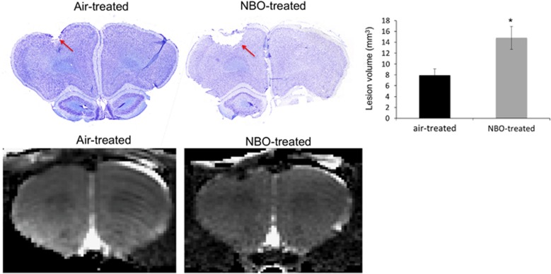
Figure 4.
Representative images of Nissl-stained brain sections from an air-treated traumatic brain injury (TBI) and normobaric oxygen therapy (NBO)-treated TBI animal on day 14 after injury with the corresponding magnetic resonance imaging (MRI) scan shown below. The lesion is indicated by the red arrows in both Nissl sections. The histogram shows the average lesion volume between air- and NBO-treated animals (mean±s.e.m., n=7 each group, *P<0.05).

