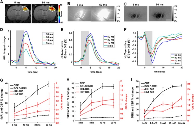
Figure 5.
Hemodynamic responses modulated by the duration of the light pulse, frequency, and power. Activation area is larger when light pulse duration increases from 5 ms (left panel) to 50 ms (right panel) in blood oxygenation level-dependent (BOLD) functional magnetic resonance imaging (fMRI) (P<0.01 and t>3) (A), deoxyhemoglobin (dHb)-weighted optical intrinsic signal (OIS) (B), and total hemoglobin (HbT)-weighted OIS (C). Scale bar in (A–C)=1 mm. The dark outline of the laser doppler flowmetry (LDF) probe can be seen in (B). Group-averaged time courses (D–F) were obtained from forelimb primary somatosensory cortex (S1FL) (n=4 for fMRI and 8 for OIS). Longer pulse duration induced larger functional signal change in BOLD fMRI (D), dHb-weighted OIS (E), and HbT-weighted OIS (F). Shaded area: the light stimulation duration of 4 seconds. Peak amplitude of LDF, BOLD fMRI, dHb-weighted OIS, and HbT-weighted OIS was plotted as a function of pulse duration (G), frequency (H), and power (I). To compare with other modalities easily, signal changes in HbT-weighted OIS were inverted to positive. Error bars: s.e.m.

