Figure 6.
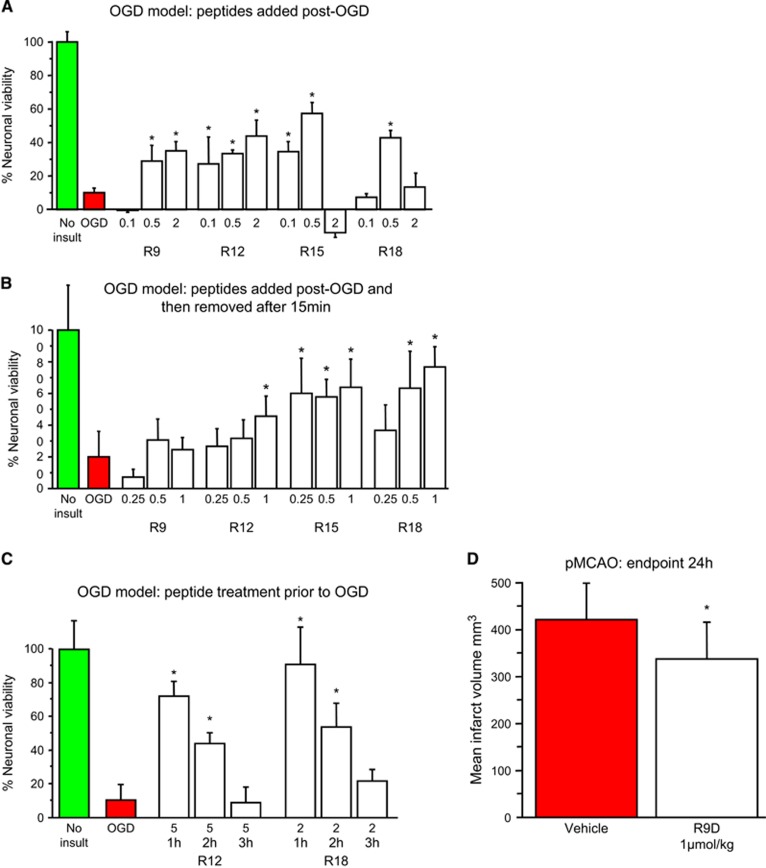
Oxygen-glucose deprivation (OGD) and permanent MCAO models. (A) Peptides added to neuronal cultures immediately after OGD. (B) Peptides added to neuronal cultures immediately after OGD and removed after 15 minutes. (C) Peptides present in neuronal cultures only for 10 minutes at 1 to 3 hours before OGD. Neuronal viability measured 20 to 24 hours after OGD. Concentration of peptide in μmol/L. MTS data were expressed as percentage neuronal viability with no insult control taken as 100% viability (mean±s.d.; n=4–6; *P<0.05). (D) Neuroprotective effects of the R9D peptide in permanent middle cerebral artery occlusion (MCAO) stroke model when administered intravenously 30 minutes after occlusion. Peptide dose was 1 μmol/kg (600 μL: intravneous) and infarct assessment was at 24 hours after MCAO (mean±s.d.; n=12; *P<0.05). Animal treatments were randomized and all procedures were performed masked to treatment, and physiologic measurements did not differ between the treatment groups (data not shown). MTS, 3-(4,5,dimethyliazol-2-yl)-5-(3-carboxymethoxy-phenyl)-2-(4-sulfophenyl)-2H-tetrazolium salt.

