Figure 3.
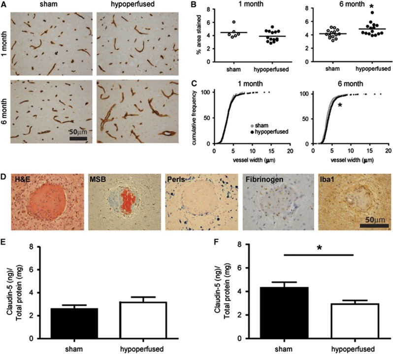
Pronounced vascular alterations and blood–brain barrier (BBB) breakdown. Collagen IV staining in sham and hypoperfused mice after 1 month (N= 6 sham; N=14 hypoperfused) or 6 months (N=14 sham; N=15 hypoperfused)(A). One-month hypoperfusion had no significant effect on density (B) or vessel width (C) in the subcortex (thalamus). In comparison, 6 months of hypoperfusion resulted in a significant increase in collagen IV density (A) and vessel width (B) within the thalamus. Evidence of fibrinoid necrosis is observed in hypoperfused mice (in 12 out of the 20) with vascular disruption (D) observed on hematoxylin and eosin (H&E) and Martius scarlet blue (MSB), with collagen accumulation, vessel wall enlargement, and fibrin deposition (red in MSB); surrounding hemosiderin blood products (Perls), parenchymal fibrogen accumulation, and inflammatory cells (ionized calcium binding adaptor molecule 1, Iba1) suggest BBB disruption and macrophage activation (features absent in 6-month sham mice, N=11). Quantification of Claudin-5 in vessel-enriched fractions by enzyme-linked Immunosorbent assay (ELISA) showed no changes in the levels of the protein after 1 month (E; N=8 sham; N=6 hypoperfused); in contrast, a significant decrease was found after 6 months in the hypoperfused group when compared with sham mice (F; N=10 sham; N=9 hypoperfused). *P<0.05.

