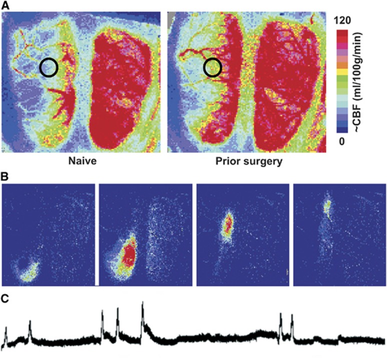
Figure 3.
Perfusion imaging of penumbral cerebral blood flow (CBF) and peri-infarct depolarization (PID)-associated hyperemic transients. (A) Distribution of CBF deficits during occlusion. Representative speckle contrast perfusion images are shown for a naive (left) and previously sham-operated animal (right), both maintained under isoflurane anesthesia during focal ischemia. Circles identify regions of interest at the margin of the middle cerebral artery (MCA) territory used for quantitative comparisons, at a location having flow values in naive animals near the perfusion threshold for infarction. Scale bar shows calibration with autoradiographic CBF derived in a previous study.5 (B) PID-associated hyperemia. Difference images obtained at 1-minute intervals illustrate the rostro-caudal migration of a PID-associated flow transient at the margin of the ischemic territory. (C) PID recording. Representative trace illustrates a series of PID-associated hyperemic events during MCA occlusion.

