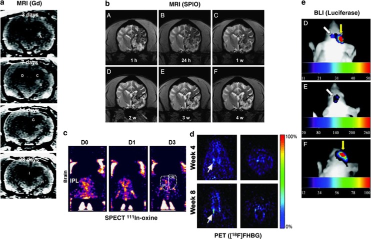
Figure 2.
Comparison of imaging techniques for transplanted therapeutic stem cells following preclinical middle cerebral artery occlusion. (A) Monitoring of Gadolinium (Gd)-labelled mesenchymal stem cells (MSCs) in rat brain using magnetic resonance imaging (MRI). White patch (arrows) shows migration of human MSCs toward the peri-infarct cortical area over 14 days.46 (B) Superparamagnetic iron oxide nanoparticle (SPIO)-labelled MSCs (area of hypointensity) viewed using magnetic resonance imaging (MRI), 1 hour to 4 weeks after transplantation into beagles.115 (C) Indium (111In)-oxine-labelled umbilical tissue-derived cells can be seen accumulating in the ipsilateral cortex (IPL. CNL=contralateral) 1 to 3 days after transplantation using single photo emission computed tomography (SPECT).76 (D) 9-(4-[18F]fluoro-3 hydroxymethylbutyl) guanine ([18F]FHBG)-labelled embryonic stem cell-derived neural stem cells (NSCs) viewed through positron emission tomography (PET) can be seen localizing in the striatal region of the forebrain.107 (E) Luciferase photon emission detected through bioluminescence imaging (BLI) 21 days after transplanting of neural progenitor cells (NPCs).88

