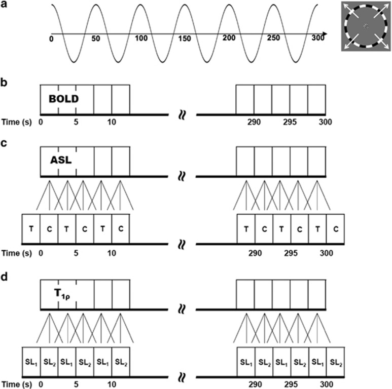
Figure 1.
Expanding-ring visual stimulus paradigm used for the functional imaging experiments. (A) Traveling wave eccentricity mapping paradigm used to activate the visual cortex. The phase of the sinusoidal wave varies as a function of distance from the center of the visual field. (B) Blood oxygenation level-dependent (BOLD) imaging experiment data acquisition. (C) The arterial spin labeling (ASL) imaging experiment calculated difference images using sliding window with linear interpolation to estimate C (control) and T (tag) images for each repetition time (TR). (D) T1ρ imaging experiment estimated the T1ρ relaxation times using a mono-exponential fit along with sliding window and linear interpolation to estimate the SL1 (spin-lock duration time=10 ms) and SL2 (spin-lock duration time=40 ms) spin-lock images for each TR.

