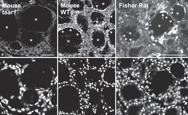
Fig. 1.
Taar1 localization in mouse and rat thyroid tissue. Cryosections through thyroid tissue obtained from taar1-/- (a), WT C57BL/6 mice (b) and Fisher rats (c) were stained with rabbit anti-mouse Taar1 polyclonal antibodies and analyzed by confocal laser scanning microscopy. Note the absence of distinct staining in taar1-deficient mouse thyroid tissue (a) and the presence of Taar1-immunoreactive structures at the apical plasma membrane (arrowheads) and within follicle cells (arrows) in WT mouse and rat thyroid glands. Asterisks indicate the follicle lumen; nuclei were counterstained with Draq5™ (a1-c1). Scale bars = 100 µm.

