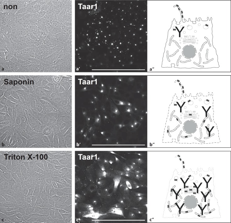
Fig. 2.
Localization of Taar1 upon differential permeabilization of FRT cells. Confocal laser scanning micrographs of formaldehyde-fixed FRT cells before (a, a') and after saponin (b, b') or Triton X-100 (c, c') permeabilization and immunolabeling with antibodies against Taar1, and corresponding schematic depictions (a“- c”) of increasing concentrations of antibodies recognizing Taar1 in intracellular compartments such as ER, Golgi apparatus and transport vesicles. Note Taar1 localization in small puncta and disc-like structures in non- and saponin-permeabilized cells (a', b'), while ER- and Golgi-localized Taar1 was detected in cells permeabilized with Triton X-100 (c'). Corresponding phase-contrast (a-c) and single-channel fluorescence microscopic images are displayed (a'–c'). Scale bars = 100 µm.

