Fig. 3.
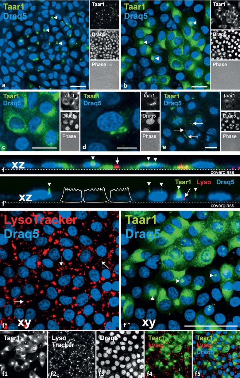
Heterogeneity of Taar1 distribution in FRT cells. Confocal laser scanning micrographs of formaldehyde-fixed and saponin-permeabilized FRT cells in confluent monolayers were immunolabeled with commercially prepared (Antibodies Online) polyclonal antibodies against Taar1 (a-f5, green signals) as well as incubated with LysoTracker Red DND-99 before fixation and immunostaining (f-f5, red signals). Immunostaining revealed the heterogeneity of the Taar1 distribution across the cell population (a, b). Note that Taar1 was found in the compartments of the secretory pathway of FRT cells, namely the ER (c), the Golgi apparatus (d) and in vesicles that lined up along the borders of neighboring cells (e, arrows). Significantly, Taar1 immunoreactivity was absent from endocytic compartments (f-f5). Taar1 was additionally present in thin and broader extensions of the apical plasma membrane domains (arrowheads), indicative of its presence in cilia. Single-channel, overlays of different channels and corresponding phase-contrast micrographs are depicted as indicated. Nuclei were counterstained with Draq5™ (blue signals). Scale bars = 20 µm (a-c, e), 10 µm (d) and 50 µm (f-f5). The images in panels c and f-f5 are extended foci of single sections, with 0°-projections along xz depicted in f and f', where the position of the cover glasses and schematic sketches of epithelial cells are given to assist with orientation.

