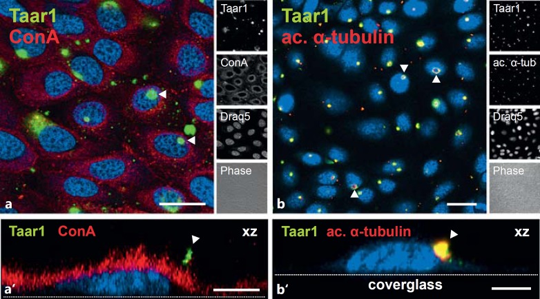
Fig. 4.
Localization of Taar1 at the surface of FRT cells. Confocal laser scanning micrographs of formaldehyde-fixed and saponin-permeabilized FRT cells in confluent monolayers were immunolabeled with commercially prepared polyclonal rabbit antibodies directed against Taar1 (green signals) and costained with the lectin ConA (a, a', red signals) or antibodies specific for acetylated α-tubulin (b, b', red signals). Lectin costaining of the glycocalyx revealed that Taar1 immunoreactivity is localized to plasma membrane appendages (arrowheads in a, a'), while antiacetylated α-tubulin staining demonstrated the presence of Taar1 in cellular extensions identified as cilia (arrowheads in b, b'). Single-channel overlays of different channels and corresponding phase-contrast micrographs are depicted as indicated. Nuclei were counterstained with Draq5™ (blue signals). Scale bars = 20 µm (a, b) and 5 µm (a', b'). The images in a' and b' are xz projections in which the positions of the cover glass are given for orientation purposes.

