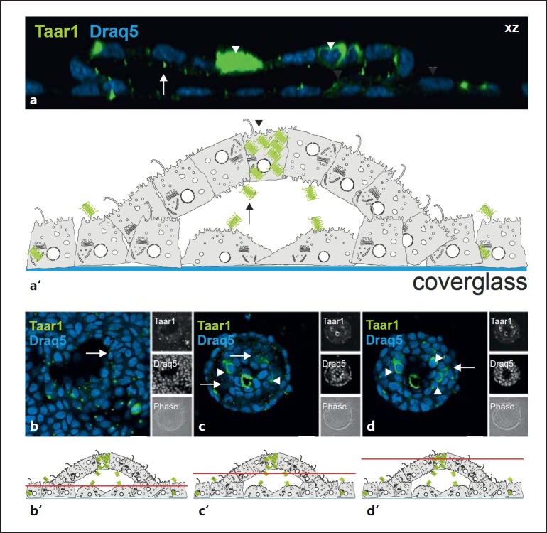
Fig. 5.
Localization of Taar1 upon formation of domes and FLS. Confocal laser scanning micrographs of formaldehyde-fixed and saponin-permeabilized FRT cells in postconfluent cultures upon formation of domes and FLS were immunolabeled with rabbit polyclonal antibodies generated against Taar1 (green signals). Side views of single sections in a z-stack are projected along the xz direction (a), single sections taken at the bottom, median and top poles of a typical FLS are depicted in xy (b-d), and corresponding schematic drawings of FLS (a') with indications on where sections were positioned in b-d (b'-d') highlight the localization of Taar1 at the apical plasma membrane domains pointing towards the inside of the forming, future FLS lumen. Note that few individual cells displayed significant expression of Taar1 (arrowheads). Single-channel and corresponding phase-contrast micrographs are depicted as indicated in the side panels. Nuclei were counterstained with Draq5™ (blue signals). Scale bars = 10 µm (a) and 20 µm (b-d).

