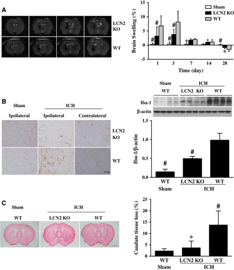
Figure 3.
(A) T2-weighted magnetic resonance imaging (MRI) images at 24 hours after 30 μL blood was injected into the right basal ganglia in lipocalin-2 knockout (LCN2−/−) or wild-type (WT) mice. Brain swelling calculated as ((ipsilateral–contralateral hemisphere)/contralateral hemisphere)) × 100% at days 1, 3, 7, 14, and 28 after blood injection in LCN2−/− or WT mice. Values are mean±s.d.; #P<0.01, *P<0.05 versus WT intracerebral hemorrhage (ICH) group. (B) Iba-1 immunoreactivity and protein levels in the basal ganglia of WT and LCN2−/− mice at 24 hours after ICH or sham operation, scale bar=50 μm. Values are mean±s.d.; n=4 for each group, #P<0.01, versus WT ICH group. (C) Ipsilateral caudate tissue loss at day 28 after 30 μL blood was injected into the right basal ganglia in LCN2−/− and WT mice. Tissue loss was calculated as ((contralateral–ipsilateral caudate)/contralateral caudate) × 100%. Values are mean±s.d.; n=4 for each group, *P<0.05, versus WT ICH group, scale bar=1 mm.

