Figure 1.
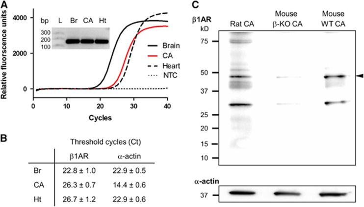
Expression of β1-adrenergic receptor (β1AR) in cerebral arteries. (A) Real-time RT-PCR amplification of β1AR cDNA from rat brain (Br), cerebral arteries (CA) and heart (Ht). Inset: Single PCR products of β1AR amplification are of predicted size (155 bp). (B) Threshold cycles (Ct) for β1AR and α-actin from three real-time PCR experiments (mean±s.e.m.). (C) Western blot immunoreactive bands corresponding to β1AR (arrowhead) in protein lysates from rat and mouse CA (30 μg/each). CA from βAR-knockout mouse (β-KO CA) and C57BL/6 J wild-type mouse (WT CA) represent negative and positive controls. Representative blot from three experiments from three separate sets of animals. L, Ladder; NTC, no template control.

