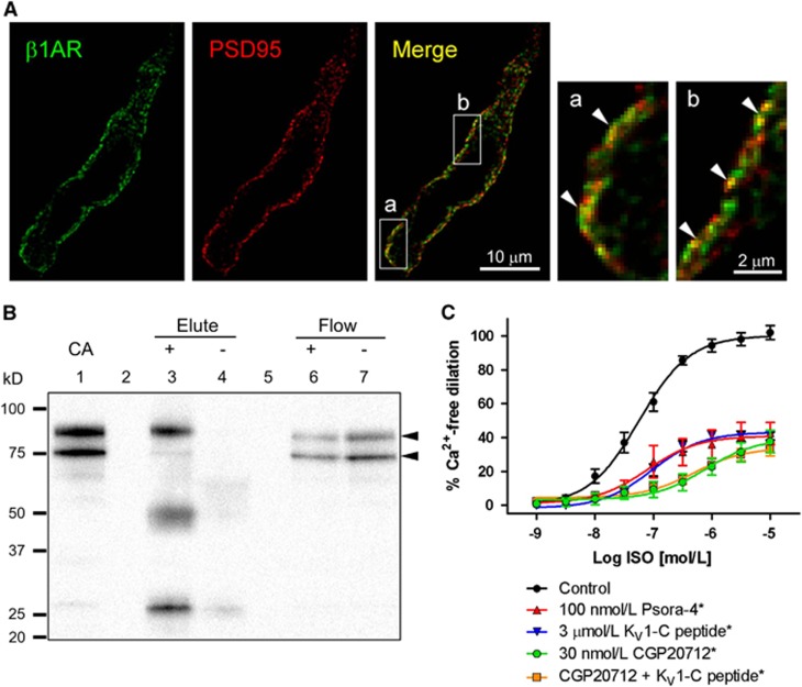
Figure 6.
Physical and functional association of β1-adrenergic receptor (β1AR) and postsynaptic density-95 (PSD95) in cerebral arteries (CA). (A) A representative confocal image of a cerebral vascular smooth muscle cell immunostained for β1AR (green) and PSD95 (red). The yellow pixels in the merged image represent colocalization. Magnified images of boxed regions (a, b) illustrate clusters of pixels with strong colocalization (arrowheads). (B) Immunoprecipitation of cerebral artery lysate using anti-β1AR antibody-conjugated bead pull-down (+). β1AR immunoprecipitate (Elute) and flow through (Flow) (lanes 3 and 6) and non-conjugated bead controls (−) (lanes 4 and 7) were probed for PSD95 (arrowheads) on a western blot. Depicted is a representative scan from three similar experiments. Lane 1: untreated cerebral artery lysate (CA). Lanes 2 and 5: empty lanes. (C) Isoproterenol (ISO) response of isolated superior cerebellar arteries (SCA) pretreated with Psora-4 (100 nmol/L), KV1-C peptide (3 μmol/L) and CGP20712 (30 nmol/L), n=5 to 6. *Significant difference from Control, P<0.05.

