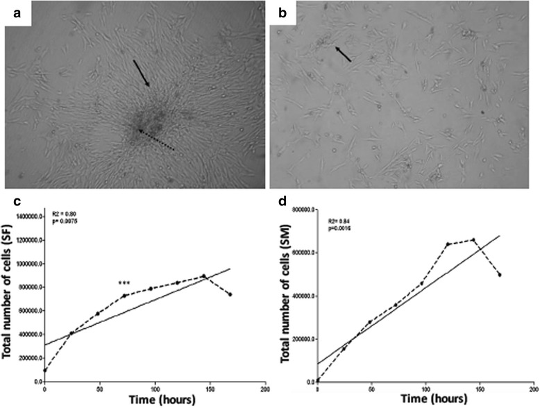
Fig. 1.

Cell morphology and growth. a and (b): Both cells, from the synovial fluid and the membrane, respectively, had a fibroblast-like appearance with a fusiform shape, growing in colonies. c and d: Both cultures had a growth pattern represented by a sigmoidal curve. The synovial fluid cells (SF) showed a period of adjustment without proliferation (lag phase, approximately 50 h of culture). Exponential growth was observed until 150 h, and after 168 h a decline phase was observed (c). In contrast for synovial membrane cells (SM) the lag phase was reached approximately 24 h of culture, followed by a exponential growth until 150 h and a decline phase close to 160 h (d)
