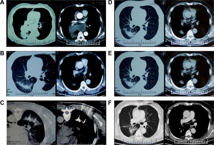
Figure 1.
CT scan images taken during the entire treatment course and follow-up.
Notes: (A) A 69-year-old male with squamous cell carcinoma at stage IIIB (T2N3M0) in the left upper lobe. (B) Target tumor regressed to a diameter of 3.8 cm after first-line treatment. (C) Double antennas were inserted simultaneously into the target tumor at prone position. (D) Non-enhanced irregular cavity showed coagulation of the target tumor without residue (complete ablation) 1 month after MWA. (E and F) The ablation zone evolved dynamically to a small fibrotic scar without local recurrence at 3 months (E) and 36 months (F) after MWA.
Abbreviations: CT, computed tomography; MWA, microwave ablation.

