
Fig. 7. TDP1 catalytic activity requirement for cellular tolerance to ABC.
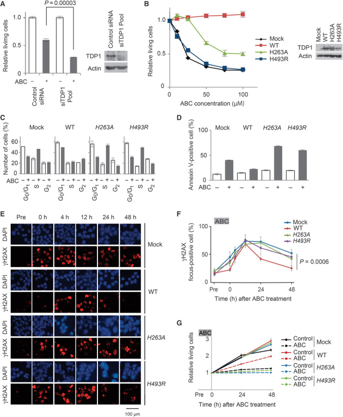
(A) Control siRNA or siTDP1 mixture was transfected into Jurkat cells. Depletion of TDP1 expression was confirmed by Western blot analysis 48 hours after transfection (right panel). Jurkat cells transfected with control siRNA or siTDP1 were treated with or without 300 μM ABC for 48 hours. MTS values of treated relative to untreated cells are shown (left panel). Results are expressed as means ± SD of three independent experiments. (B) MT-2 cells stably transfected with either wild-type (WT) TDP1, H263A TDP1, or H493R TDP1 transgenes. Stably transfected clones and control cells (mock) were treated with the indicated dose of ABC for 48 hours. Western blot analysis was conducted for expression of transgenes (right panel). Data shown as in (A, left panel). (C) Cell cycle analysis is shown as in Fig. 2A. MT-2/TDP1WT, MT-2/TDP1H263A, and MT-2/TDP1H493R cells were treated with or without 100 μM ABC for 24 hours and subjected to flow cytometry. (D) The extent of apoptosis is shown as the percentage of annexin V–positive cells (y axis). Cells were treated as in (C). (E) The indicated cells were treated as in Fig. 3C. Data shown as in Fig. 3C. (F) Statistical analysis of data in (E). MT-2/TDP1WT cells, but not MT-2/TDP1H263A or MT-2/TDP1H493R cells, are resistant to ABC. (G) Proliferation of the indicated cells is shown as in Fig. 3E.