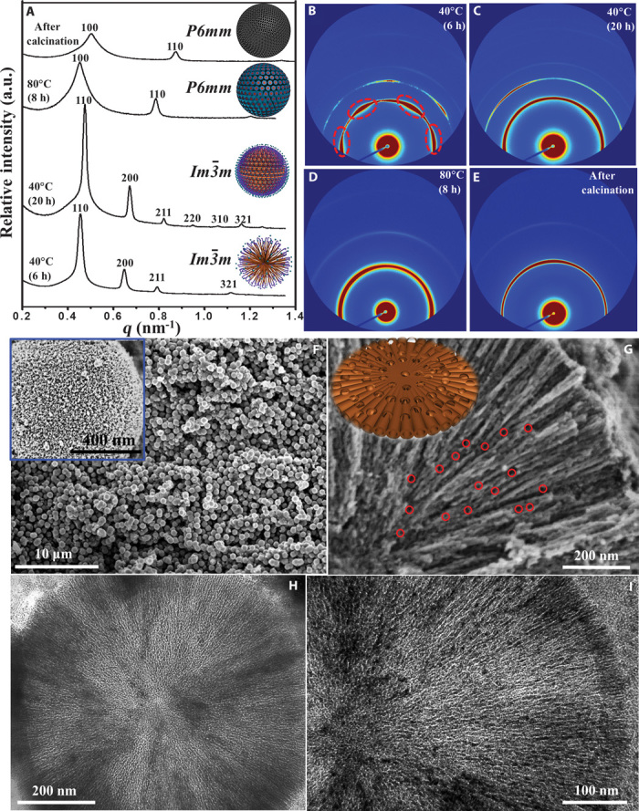
Fig. 2. Microstructure characterization of the radially oriented mesoporous TiO2 microspheres.

(A) In situ synchrotron radiation 1D SAXS patterns of the mesoporous TiO2 microsphere products harvested at different intervals of reaction time. Insets: Corresponding schematic representation of the four samples. a.u., arbitrary units. (B to E) 2D SAXS images of the four samples. (F) SEM image of the mesoporous TiO2 microspheres. Inset: SEM image of a single mesoporous TiO2 microsphere. (G) SEM image of a single ultramicrotomed, radially-oriented mesoporous TiO2 microsphere with a large number of interchannel pores (~5 to 15 nm in diameter, marked by red circles). Inset: Corresponding schematic representation of the structure models for the radially oriented channels with interchannel pores. (H and I) TEM images of a single ultramicrotomed, mesoporous TiO2 microsphere.
