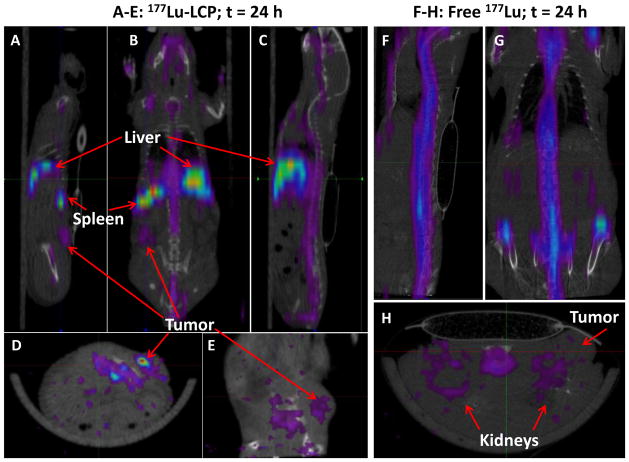
Figure 5.
SPECT/CT images of nude mouse bearing subcutaneous UMUC3/3T3 tumor on left flank at 24 h after injection of 177Lu-LCP (Figures 5A–E) or free 177Lu (Figures 5F–H). Mouse slit collimator was used for whole body imaging: A) left sagittal, B) coronal, C) midsagittal, F) mid-sagittal and G) coronal views. Pinhole collimator was used in D), E), and H) for higher resolution axial and left sagittal views. 177Lu-LCP accumulated in tumor, liver, and spleen, while free 177Lu accumulated in the bone and kidneys, but not in the tumor.

