
Fig. 1.
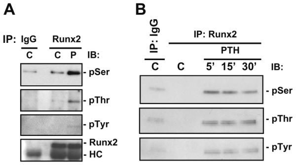
PTH stimulation of Runx2 phosphorylation in vivo. (A) UMR 106-01 cells were treated with control or PTH (10−8M)-containing media for 5 min. (B) UMR 106-01 cells were treated with control or PTH (10−8M)-containing media for 5, 15 and 30 min. Whole cell lysates were prepared and immunoprecipitated with either IgG or Runx2 antibody and immunoblotted with antibodies as indicated followed by detection by ECL kit (C, control; P, PTH; IP, immunoprecipitation; IB, immunoblot; HC, heavy chain; p, phosphorylated).