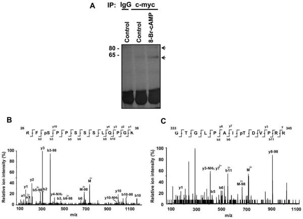
Fig. 3.
MALDI/MS analysis of Runx2 phosphorylation sites in vivo. (A) The c-myc-tagged Runx2 construct (pCMV–c-myc–Runx2) was transiently transfected into COS-7 cells. The cells were then treated with control or 8-Br-cAMP (10−3M)-containing media for 5 min. Whole cell lysates were prepared, immunoprecipitated with antibody to the c-myc tag, fractionated by 12% SDS–PAGE and silver stained. The two protein bands corresponding to 65 kDa and 80 kDa were excised from the gel and subjected to MALDI/MS analysis. (B) MS/MS spectrum of a doubly-charged ion corresponds to RFSPPSSSLQPGK plus one phosphorylation site (m/z 734.42). The observed y- and b-ion series confirmed the peptide sequence and localized the phosphorylation site at S28. (C) MS/MS spectrum of a doubly-charged ion corresponds to GTGLPAITDVPRR plus one phosphorylation site (m/z 716.87). The observed y- and b-ion series tentatively confirmed the peptide sequence and localized the phosphorylation site at T340.

