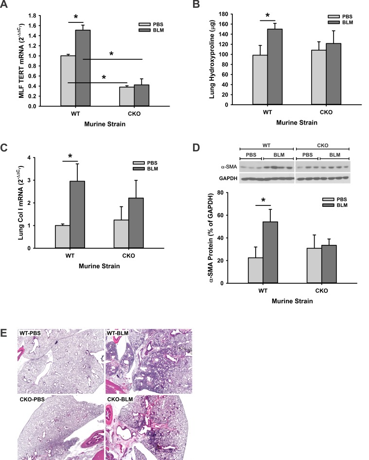
Fig 5. The impairment of pulmonary fibrosis in TERT CKO mice.
(A) The MLF were isolated from TERT CKO or WT mice at 21 days after BLM injection, and analyzed for TERT mRNA by qRT-PCR. The expression was expressed as the fold change of the level in PBS-treated WT MLF. n = 3–5 mice per group. (B) The lungs from TERT CKO and control mice were homogenized at day 21 after BLM treatment, and measured for whole lung collagen content by HYP assay. n = 3–5 mice per group. (C) Lung tissue RNA extracted from the indicated murine strains was also analyzed for type I collagen mRNA by qRT-PCR. n = 3–5 mice per group. (D) Lung tissue lysates were prepared by RIPA buffer, and analyzed for α-SMA protein in the indicated murine strain by Western blotting (top panel). Quantitative data was normalized by the internal control GAPDH, and shown as the percentage of the GAPDH signals (bottom panel). n = 3–4 mice per group. (E) Representative H & E stained lung tissue sections at day 21after BLM treatment are shown. Original magnification × 20. *, P < 0.05.

