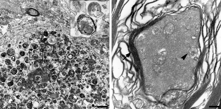
Fig 4. Transmission electron microscopy of spinal cord spheroids compared to a normally structured axon.
Ultrastructurally, spheroids lacked myelin sheaths and contained closely packed accumulations of membrane-bound vacuolar structures. High numbers of vacuoles were rimmed by a double layered membrane separated by an electron-lucent cleft and defined as autophagosomes (insert; A). The axons of a healthy, age matched Beagle dog is surrounded by a thick myelin sheath (arrowhead). Isolated vacuolar structures, interpreted as mitochondria (arrow) are present between neurofilaments (B). (A) Bar: overview: 1 μm; insert 0.5 μm. (B) Bar: 2.3 μm.

