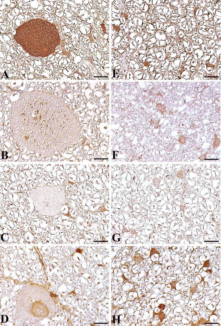
Fig 5. Expression of autophagosome, lysosome and late endosome associated proteins in the spinal cord white matter of an NAD affected Spanish water dog (A-D) and an age matched control Beagle dog (E-H).
(A) In the affected Spanish water dog, immunohistochemistry using a LC3B specific antibody demonstrated an autophagosome accumulation within spheroids. Only few LAMP2 positive lysosomes (B) and RAB7 stained late endosomes (C) are present within spheroids. No TECPR2 accumulations are found within spheroids (D). In diseased Spanish water dogs, LC3B, LAMP2 and TECPR2 are strongly expressed in glial cells and axons without spheroid formation. In the age matched healthy Beagle dog, LC3B (E), LAMP2 (F) and TECPR2 (H) positive staining is found in axons and glial cells. RAB 7 (G) positive staining is restricted to glial cells. Bar: 20 μm. A-H: Nomarski differential interference-contrast optic.

