Fig. 2.
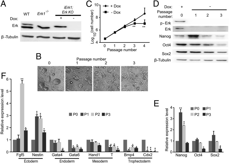
Erk signaling is essential for the self-renewal of ESCs. (A) Western blots demonstrate the diminishment of Erk expression in iErk1; Erk KO ESCs at 48 h after Dox withdrawal. (B) Colony morphology change upon Erk KO. iErk1; Erk KO ESCs were continuously cultured in the absence of Dox for three passages. Phase-contrast images of ESC colonies at each passage are shown. (C) Reduced proliferation of ESCs after Erk KO. iErk1; Erk KO ESCs were cultured in serum/LIF medium with or without Dox. Cell numbers were counted every passage, and equal amounts of ESCs were plated onto tissue-culture dishes. (D–F) Expression dynamics of pluripotency and differentiation markers after Erk KO. Cells were cultured as described in C. The protein (D) and mRNA (E) levels of pluripotency markers Nanog, Oct4, and Sox2 were measured by Western blot and quantitative RT-PCR. (F) Expression of differentiation genes after Erk KO. *P < 0.05; **P < 0.01. Error bars are SDs.

